What is the Normal Anatomy of the Foot and Ankle?
The foot and ankle form complex joints that are involved in movement and providing stability and balance to the body. The foot and ankle consist of 26 bones, 33 joints, and many muscles, tendons, and ligaments.
Bones of the Ankle
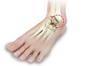
The ankle joint connects the leg with the foot and is composed of three bones: the tibia, fibula, and talus. The tibia or shinbone and fibula or calf bone are bones of the lower leg, which articulate with the talus or ankle bone, enabling up and down movement of the foot.
Three bony bumps present on the ends of the tibia and fibula form parts of the ankle joint:
- The medial malleolus, formed by the tibia, is found on the inside of the ankle.
- The posterior malleolus, also formed by the tibia, is found at the back of the ankle.
- The lateral malleolus, formed by the fibula, is found on the outer aspect of the ankle.
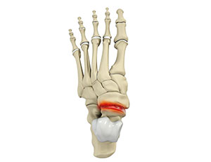
Bones of the Feet
The foot acts as a single functional unit, but can be divided into three parts: the hindfoot, midfoot and forefoot.
The hindfoot forms the ankle and heel, and is made up of the talus bone and calcaneus or heel bone. The heel bone is the largest bone in the foot.
The midfoot connects the hindfoot to the forefoot, and consists of one navicular bone, one cuboid bone, and three cuneiform bones. The navicular bone is found in front of the heel bone, and the cuneiform and cuboid bones are arranged in front of the navicular bone.
These bones are connected to five metatarsal bones of the forefoot that form the arch of the foot for shock absorption while walking or running. The forefoot is also made up of the toes or digits, formed by bones called phalanges - three in each toe, except the big toe, which has only two phalanges. The big toe has two additional tiny round sesamoid bones in the ball of the foot, which helps in upward and downward movements of the toe.
Ankle and Foot Joints
There are 33 joints in the ankle and foot. They include:
- Hinge joints in the ankle, which allow flexion (bending) and extension
- Gliding joints found in the hindfoot, which allow gliding movements
- Condyloid joints found in the forefoot and toes, which allow the flexion (bending) and extension, adduction, and abduction (side-waze movement).
The joints of the foot and ankle provide stability and support the weight of your body, helping you to walk or run, and adapt to uneven grounds.
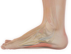
Soft Tissues of the Ankle and Foot
Our feet and ankle bones are held in place and supported by various soft tissues such as cartilage, ligaments, muscles, tendons, and bursae.
The joint surface of all the bones of the ankle and foot are lined by a thin, tough, flexible, and slippery surface called the articular cartilage, which acts as a shock absorber and cushion to reduce friction between the bones. The cartilage is lubricated by synovial fluid, which further enables smooth movement of the bones.
Ligaments are tough rope-like tissue that connect bones to other bones, and hold them in place, providing stability to the joints. The plantar fascia is the largest ligament in the foot, originating from the heel bone to the forefoot, it extends along the lower side of the foot and is involved in maintaining the arch of the foot. The plantar fascia ligament stretches and contracts to provide balance and strength to the foot. Lateral ligaments on the outside of the foot and medial ligaments on the inside of the foot provide stability and allow up and down movement of the foot.
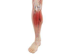
The foot is made up of 20 muscles that help in movement. The main muscles include:
- Anterior tibial muscle, which allows up and down movement of the foot
- Posterior tibial muscle, which supports the arch
- Peroneal tibial muscle, which controls movement on the outside of the ankle
- Extensors, which enable the ankle to raise the toes just before stepping forward
- Flexors, which stabilize the toes against the floor
- Smaller muscles that help the toes to lift and curl
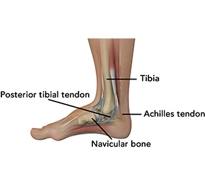
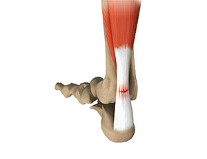
Tendons are soft tissues that connect muscles to bones. The largest and strongest tendon in the foot is the Achilles tendon, present at the back of the lower leg around the heel bone. Other tendons include peroneal and anterior and posterior tibialis.
Bursae are small fluid-filled sacs that decrease friction between tendons and bone or skin. They contain special cells called synovial cells that secrete a lubricating fluid.
-
Ankle Arthroscopy
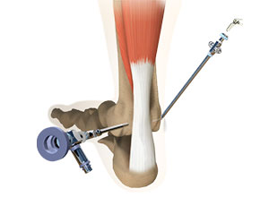
Ankle arthroscopy is a minimally invasive surgical procedure in which an arthroscope, a small, soft, flexible tube with a light and video camera at the end, is inserted into the ankle joint to evaluate and treat a variety of conditions. The camera projects an image of the inside of the joint onto a large monitor, allowing your surgeon to look for any damage, assess the type of injury and repair the problem.
-
Arthroscopic Ankle Joint Cartilage Repair
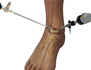
Arthroscopic ankle joint cartilage repair is a minimally invasive surgical procedure designed to repair damaged cartilage within the ankle joint using arthroscopic guidance and small incisions. The procedure enables your physician to detect the damaged cartilage and make repairs to your ankle without making large cuts in the skin and tissue as in an open ankle surgery.
-
BioCartilage Regeneration
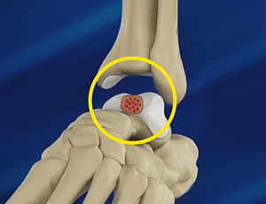
BioCartilage regeneration is a surgical technique to induce cartilage regeneration in areas of articular cartilage defects, typically in a joint such as a foot and ankle. BioCartilage contains the extracellular matrix that is native to articular cartilage including key components such as type II collagen, proteoglycans, and additional cartilaginous growth factors that stimulate growth and healing in the cartilaginous cells.
-
Ankle Ligament Reconstruction
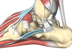
Ankle ligament reconstruction is a surgical procedure typically performed to treat serious sprains or instability in the ankle.
-
Achilles Tendon Repair
Tendons are the soft tissues connecting muscle to bone. The Achilles tendon is the longest tendon in the body and is present behind the ankle, joining the calf muscles with the heel bone. Contraction of the calf muscles tightens the Achilles tendon and pulls the heel, enabling the foot and toe movements necessary for walking, running and jumping.
-
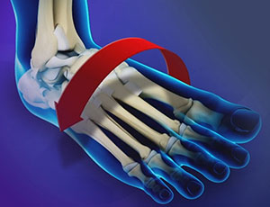
Foot and Ankle Deformity Correction
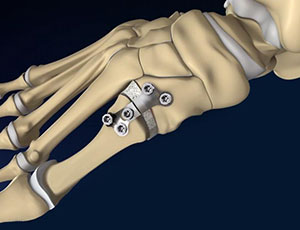
Foot and ankle deformity is the structural abnormality caused by misalignment of the bones of the foot and ankle.
-
Ankle Joint Replacement
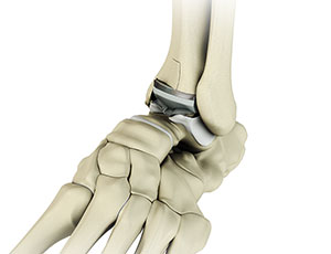
Ankle joint replacement, also known as total ankle arthroplasty, is a surgical procedure performed to relieve pain and immobility due to severe end-stage arthritis that has not responded to non-surgical treatments. The goal of ankle joint replacement surgery is to eliminate your pain and increase the mobility of your ankle joint.
-
Treatment of Foot and Ankle Sports Injuries

Injuries during sports are common. They can result from accidents, inadequate training, improper use of protective devices, or insufficient stretching or warm-up exercises. Injuries to the foot and ankle are common while playing sports such as football, hockey, skating and in weekend athletes.
-
Ankle Arthrodesis
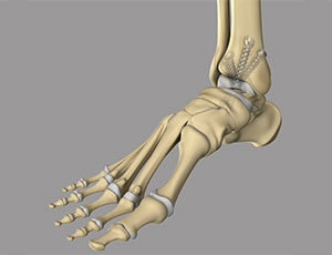
Ankle arthrodesis is the surgical fusion of bones that form the ankle joint. The ankle joint is formed by the tibia, talus, and the fibula bones. The goal of ankle arthrodesis is to relieve pain in the affected joint. This is achieved by surgically eliminating the joint.
-
Hindfoot Reconstructive Procedures
Hindfoot reconstructive procedures are a group of advanced surgical techniques used to restore alignment, stability, and function of the hindfoot, which includes the heel (calcaneus), ankle, and surrounding joints.
-
Ankle Sprain
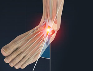
A sprain is the stretching or tearing of ligaments. Ligaments connect adjacent bones and provide stability to a joint. An ankle sprain is a common injury that occurs when you suddenly fall or twist the ankle joint, or when you land your foot in an awkward position after a jump. Most commonly, it occurs when you participate in sports, or jump or run on a surface that is irregular.
-
Ankle Instability
The joints of the ankle are held in place and stabilized by strong bands of tissue called ligaments. Ankle instability is a chronic condition characterized by a recurrent slipping of the outer side of the ankle. It usually results from repeated ankle sprains, which are injuries to the ligaments. Ankle instability is generally noticed when you move your ankle joint but can also occur while standing.
-
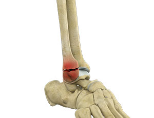
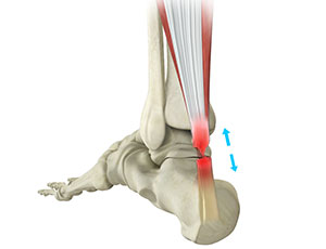
Achilles Tendon Rupture
The Achilles tendon is a strong fibrous cord present behind the ankle that connects the calf muscles to the heel bone. It is used when you walk, run and jump. The Achilles tendon ruptures most often in athletes participating in sports that involve running, pivoting and jumping. Recreational sports that may cause Achilles rupture include tennis, football, basketball, and gymnastics.
-
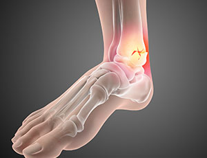
Foot and Ankle Arthritis
Arthritis is the inflammation of joints as a result of degeneration of the smooth cartilage that lines the ends of bones in a joint. This degeneration of the cartilages leads to painful rubbing of the bones, swelling, and stiffness in the joints, resulting in restricted movements.
-
Hindfoot Arthritis
Hindfoot arthritis refers to arthritis that affects the joints and structures in the hindfoot region of the foot. The hindfoot includes the talus bone, calcaneus (heel bone), and the joints connecting them, such as the subtalar joint and the talonavicular joint.
-
Shin Splints
Shin splints or medial tibial stress syndrome (MTSS) is pain around the tibia or shinbone due to inflammation of the tendons, muscles and bone tissue. It occurs because of vigorous exercises and sports activities.
-
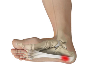
Heel Pain
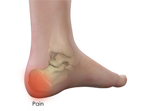
The heel is made up of the calcaneus bone and supported by a network of muscles, tendons, ligaments and soft tissues, which together support the weight of the body and stress during movement. Heel pain is a common symptom of excessive strain placed on the structures that form the heel.
-
Stress Fractures of Foot and Ankle
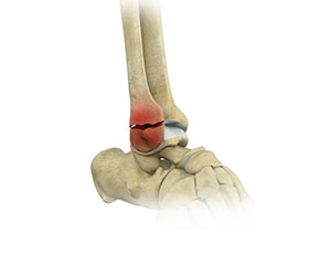
A stress fracture is described as a small crack in the bone which occurs from an overuse injury of a bone. It commonly develops in the weight-bearing bones of the lower leg and foot. When the muscles of the foot are overworked or stressed, they are unable to absorb the stress and when this happens the muscles transfer the stress to the bone which results in stress fracture.
-
Lisfranc (Midfoot) Injury
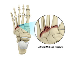
The tarsometatarsal joint or Lisfranc joint is the region in the middle of the foot formed by the articulation of the tarsal bones (a cluster of seven bones) and metatarsal bones (a group of five long bones). This region supports the arch of the foot. Lisfranc or midfoot fractures are breaks in the bones of the midfoot.
-
Tarsal Coalition
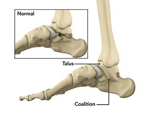
Tarsal coalition is a foot disorder that occurs due to an abnormal connection between two or more tarsal bones. These bones are located at the back of the foot and in the heel. Basically, with this condition, the bones tend to grow within one another and are joined together with the help of surrounding bones and cartilage.